图书简介
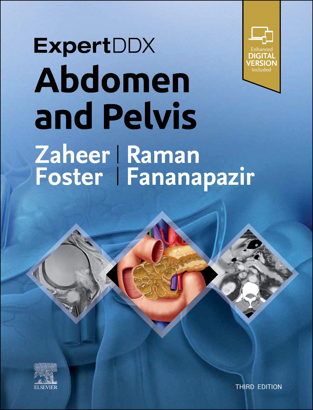
Highly practical and user-friendly, ExpertDDx: Abdomen and Pelvis, third edition, helps you reach accurate, clinically useful differential diagnoses in your everyday practice. It presents the most useful differential diagnoses for each region of the abdomen and pelvis, grouped according to anatomic location, generic imaging findings, modality-specific findings, or clinical-based indications. Each differential diagnosis includes several high-quality, succinctly annotated images; a list of diagnostic possibilities sorted as common, less common, and rare but important; and brief, bulleted text offering helpful diagnostic clues. It’s an excellent resource for subspecialty abdominal imagers as well as general radiologists and trainees, providing invaluable assistance in reaching logical, on-target differential diagnoses based on key imaging findings and clinical details. Covers 175 of the most common diagnostic challenges in abdominal and pelvic imaging, enhanced by more than 2,100 radiologic images, full-color illustrations, clinical and histologic photographs, and gross pathology images Provides a quick review of the salient features of each entity, differentiating features from other similar-appearing abnormalities Includes new chapters on hematuria, flank pain, acute scrotal pain, and seminal vesicle Adds greater focus to advancing prostate imaging methods with expanded content on lesions in the peripheral zone and lesions in the transition zone, as well as new coverage of transplant imaging Contains updates to numerous classifications, including LI-RADS for liver, O-RADS for ovarian masses, and the Tanaka classification for pancreatic cysts Features new MR examples and MR-specific diagnoses throughout, plus new differentials for contrast-enhanced ultrasound findings related to liver and kidney lesions Includes the enhanced eBook version, which allows you to search all text, figures, and references on a variety of devices
SECTION 1: PERITONEUM AND MESENTERY GENERIC IMAGING PATTERNS 4 Mesenteric or Omental Mass (Solid) Siva P. Raman, MD 10 Mesenteric or Omental Mass (Cystic) Siva P. Raman, MD 14 Fat-Containing Lesion, Peritoneal Cavity Siva P. Raman, MD 18 Mesenteric Lymphadenopathy Siva P. Raman, MD 22 Abdominal Calcifications Siva P. Raman, MD 28 Pneumoperitoneum Siva P. Raman, MD 32 Hemoperitoneum Siva P. Raman, MD 36 Misty (Infiltrated) Mesentery Siva P. Raman, MD MODALITY-SPECIFIC IMAGING FINDINGS COMPUTED TOMOGRAPHY 42 High-Attenuation (Hyperdense) Ascites Siva P. Raman, MD SECTION 2: ABDOMINAL WALL ANATOMICALLY BASED DIFFERENTIALS 48 Abdominal Wall Mass Siva P. Raman, MD 52 Mass in Iliopsoas Compartment Siva P. Raman, MD 54 Groin Mass Siva P. Raman, MD 58 Elevated or Deformed Hemidiaphragm Siva P. Raman, MD 60 Defect in Abdominal Wall (Hernia) Siva P. Raman, MD SECTION 3: ESOPHAGUS GENERIC IMAGING PATTERNS 66 Intraluminal Mass, Esophagus Atif Zaheer, MD and Michael P. Federle, MD, FACR 68 Extrinsic Mass, Esophagus Atif Zaheer, MD and Michael P. Federle, MD, FACR 72 Lesion at Pharyngoesophageal Junction Atif Zaheer, MD and Michael P. Federle, MD, FACR 74 Esophageal Ulceration Atif Zaheer, MD and Michael P. Federle, MD, FACR 76 Mucosal Nodularity, Esophagus Atif Zaheer, MD and Michael P. Federle, MD, FACR 78 Esophageal Strictures Atif Zaheer, MD and Michael P. Federle, MD, FACR 80 Dilated Esophagus Atif Zaheer, MD and Michael P. Federle, MD, FACR 82 Esophageal Outpouchings (Diverticula) Atif Zaheer, MD and Michael P. Federle, MD, FACR 84 Esophageal Dysmotility Atif Zaheer, MD and Michael P. Federle, MD, FACR CLINICALLY BASED DIFFERENTIALS 86 Odynophagia Atif Zaheer, MD and Michael P. Federle, MD, FACR SECTION 4: STOMACH GENERIC IMAGING PATTERNS 90 Gastric Mass Lesions Atif Zaheer, MD and Michael P. Federle, MD, FACR 96 Intramural Mass, Stomach Atif Zaheer, MD and Michael P. Federle, MD, FACR 98 Target or Bull’s-Eye Lesions, Stomach Atif Zaheer, MD and Michael P. Federle, MD, FACR 100 Gastric Ulceration (Without Mass) Atif Zaheer, MD and Michael P. Federle, MD, FACR 102 Intrathoracic Stomach Atif Zaheer, MD and Michael P. Federle, MD, FACR 104 Thickened Gastric Folds Atif Zaheer, MD and Michael P. Federle, MD, FACR 110 Gastric Dilation or Outlet Obstruction Atif Zaheer, MD and Michael P. Federle, MD, FACR 114 Linitis Plastica, Limited Distensibility Atif Zaheer, MD and Michael P. Federle, MD, FACR CLINICALLY BASED DIFFERENTIALS 118 Epigastric Pain Atif Zaheer, MD and Michael P. Federle, MD, FACR 124 Left Upper Quadrant Mass Atif Zaheer, MD and Michael P. Federle, MD, FACR SECTION 5: DUODENUM GENERIC IMAGING PATTERNS 130 Duodenal Mass Atif Zaheer, MD and Michael P. Federle, MD, FACR 136 Dilated Duodenum Atif Zaheer, MD and Michael P. Federle, MD, FACR 138 Thickened Duodenal Folds Atif Zaheer, MD and Michael P. Federle, MD, FACR SECTION 6: SMALL INTESTINE GENERIC IMAGING PATTERNS 142 Multiple Masses or Filling Defects, Small Bowel Atif Zaheer, MD and Michael P. Federle, MD, FACR 144 Cluster of Dilated Small Bowel Atif Zaheer, MD and Michael P. Federle, MD, FACR 146 Aneurysmal Dilation of Small Bowel Lumen Atif Zaheer, MD and Michael P. Federle, MD, FACR 148 Stenosis, Terminal Ileum Atif Zaheer, MD and Michael P. Federle, MD, FACR 150 Segmental or Diffuse Small Bowel Wall Thickening Atif Zaheer, MD and Michael P. Federle, MD, FACR 156 Pneumatosis of Small Intestine or Colon Atif Zaheer, MD and Michael P. Federle, MD, FACR CLINICALLY BASED DIFFERENTIALS 160 Occult GI Bleeding Atif Zaheer, MD and Michael P. Federle, MD, FACR 164 Small Bowel Obstruction Atif Zaheer, MD and Michael P. Federle, MD, FACR SECTION 7: COLON GENERIC IMAGING PATTERNS 172 Solitary Colonic Filling Defect Atif Zaheer, MD and Michael P. Federle, MD, FACR 174 Multiple Colonic Filling Defects Atif Zaheer, MD and Michael P. Federle, MD, FACR 176 Mass or Inflammation of Ileocecal Area Atif Zaheer, MD and Michael P. Federle, MD, FACR 182 Colonic Ileus or Dilation Atif Zaheer, MD and Michael P. Federle, MD, FACR 186 Toxic Megacolon Atif Zaheer, MD and Michael P. Federle, MD, FACR 188 Rectal or Colonic Fistula Atif Zaheer, MD and Michael P. Federle, MD, FACR 194 Segmental Colonic Narrowing Atif Zaheer, MD and Michael P. Federle, MD, FACR 198 Colonic Thumbprinting Atif Zaheer, MD and Michael P. Federle, MD, FACR 200 Colonic Wall Thickening Atif Zaheer, MD and Michael P. Federle, MD, FACR 206 Smooth Ahaustral Colon Atif Zaheer, MD and Michael P. Federle, MD, FACR CLINICALLY BASED DIFFERENTIALS 208 Acute Right Lower Quadrant Pain Atif Zaheer, MD and Michael P. Federle, MD, FACR 214 Acute Left Lower Quadrant Pain Atif Zaheer, MD and Michael P. Federle, MD, FACR SECTION 8: SPLEEN GENERIC IMAGING PATTERNS 222 Splenomegaly Siva P. Raman, MD 226 Multiple Splenic Calcifications Siva P. Raman, MD 228 Solid Splenic Mass or Masses Siva P. Raman, MD 230 Cystic Splenic Mass Siva P. Raman, MD MODALITY-SPECIFIC IMAGING FINDINGS COMPUTED TOMOGRAPHY 232 Diffuse Increased Attenuation, Spleen Siva P. Raman, MD SECTION 9: LIVER GENERIC IMAGING PATTERNS 236 Liver Mass With Central or Eccentric Scar Atif Zaheer, MD and Michael P. Federle, MD, FACR 240 Focal Liver Lesion With Hemorrhage Atif Zaheer, MD and Michael P. Federle, MD, FACR 244 Liver "Mass" With Capsular Retraction Atif Zaheer, MD and Michael P. Federle, MD, FACR 246 Fat-Containing Liver Mass Atif Zaheer, MD and Michael P. Federle, MD, FACR 248 Cystic Hepatic Mass Atif Zaheer, MD and Michael P. Federle, MD, FACR 252 Focal Hypervascular Liver Lesion Atif Zaheer, MD and Michael P. Federle, MD, FACR 258 Liver Mass With Mosaic Enhancement Atif Zaheer, MD 262 Mosaic or Patchy Hepatogram Atif Zaheer, MD and Michael P. Federle, MD, FACR 266 Hepatic Calcifications Atif Zaheer, MD and Michael P. Federle, MD, FACR 270 Liver Lesion Containing Gas Atif Zaheer, MD and Michael P. Federle, MD, FACR 274 Portal Venous Gas Atif Zaheer, MD and Michael P. Federle, MD, FACR 276 Widened Hepatic Fissures Atif Zaheer, MD and Michael P. Federle, MD, FACR 278 Dysmorphic Liver With Abnormal Bile Ducts Atif Zaheer, MD and Michael P. Federle, MD, FACR 282 Focal Hyperperfusion Abnormality (THAD or THID) Atif Zaheer, MD and Michael P. Federle, MD, FACR MODALITY-SPECIFIC IMAGING FINDINGS MAGNETIC RESONANCE IMAGING 288 Multiple Hypodense Liver Lesions Atif Zaheer, MD and Michael P. Federle, MD, FACR 294 Multiple Hypointense Liver Lesions (T2WI) Atif Zaheer, MD and Michael P. Federle, MD, FACR 298 Hyperintense Liver Lesions (T1WI) Atif Zaheer, MD and Michael P. Federle, MD, FACR 304 Liver Lesion With Capsule or Halo on MR Atif Zaheer, MD and Michael P. Federle, MD, FACR COMPUTED TOMOGRAPHY 308 Focal Hyperdense Hepatic Mass on Nonenhanced CT Atif Zaheer, MD and Michael P. Federle, MD, FACR 312 Periportal Lucency or Edema Atif Zaheer, MD and Michael P. Federle, MD, FACR 318 Widespread Low Attenuation Within Liver Atif Zaheer, MD and Michael P. Federle, MD, FACR ULTRASOUND 322 Focal Hepatic Echogenic Lesion /- Acoustic Shadowing Atif Zaheer, MD, Gregory E. Antonio, MD, DRANZCR, FHKCR, and Eric K. H. Liu, PhD, RDMS 328 Hyperechoic Liver, Diffuse Atif Zaheer, MD, Gregory E. Antonio, MD, DRANZCR, FHKCR, and Eric K. H. Liu, PhD, RDMS 330 Hepatomegaly Hee Sun Park, MD, PhD and Aya Kamaya, MD, FSRU, FSAR 334 Diffuse Liver Disease Hee Sun Park, MD, PhD and Aya Kamaya, MD, FSRU, FSAR 336 Cystic Liver Lesion Hee Sun Park, MD, PhD and Aya Kamaya, MD, FSRU, FSAR 340 Hypoechoic Liver Mass Hee Sun Park, MD, PhD and Aya Kamaya, MD, FSRU, FSAR 344 Echogenic Liver Mass Hee Sun Park, MD, PhD and Aya Kamaya, MD, FSRU, FSAR 348 Target Lesions in Liver Hee Sun Park, MD, PhD and Aya Kamaya, MD, FSRU, FSAR 350 Multiple Hepatic Masses Hee Sun Park, MD, PhD and Aya Kamaya, MD, FSRU, FSAR 354 Hepatic Mass With Central Scar Hee Sun Park, MD, PhD and Aya Kamaya, MD, FSRU, FSAR 356 Periportal Lesion Hee Sun Park, MD, PhD and Aya Kamaya, MD, FSRU, FSAR 360 Irregular Hepatic Surface Aya Kamaya, MD, FSRU, FSAR 362 Portal Vein Abnormality Hee Sun Park, MD, PhD and Aya Kamaya, MD, FSRU, FSAR SECTION 10: GALLBLADDER GENERIC IMAGING PATTERNS 366 Distended Gallbladder Siva P. Raman, MD 368 Gas in Bile Ducts or Gallbladder Siva P. Raman, MD 372 Focal Gallbladder Wall Thickening Siva P. Raman, MD 374 Diffuse Gallbladder Wall Thickening Jade Wong-You-Cheong, MBChB, MRCP, FRCR, FSRU, FSAR MODALITY-SPECIFIC IMAGING FINDINGS COMPUTED TOMOGRAPHY 378 High-Attenuation (Hyperdense) Bile in Gallbladder Siva P. Raman, MD ULTRASOUND 380 Hyperechoic Gallbladder Wall Jade Wong-You-Cheong, MBChB, MRCP, FRCR, FSRU, FSAR 382 Echogenic Material in Gallbladder Jade Wong-You-Cheong, MBChB, MRCP, FRCR, FSRU, FSAR 384 Dilated Gallbladder Jade Wong-You-Cheong, MBChB, MRCP, FRCR, FSRU, FSAR 388 Intrahepatic and Extrahepatic Duct Dilatation L. Nayeli Morimoto, MD and Aya Kamaya, MD, FSRU, FSAR CLINICALLY BASED DIFFERENTIALS 390 Right Upper Quadrant Pain Siva P. Raman, MD SECTION 11: BILIARY TRACT GENERIC IMAGING PATTERNS 398 Dilated Common Bile Duct Siva P. Raman, MD 404 Asymmetric Dilation of Intrahepatic Bile Ducts Siva P. Raman, MD 408 Biliary Strictures, Multiple Siva P. Raman, MD MODALITY-SPECIFIC IMAGING FINDINGS MAGNETIC RESONANCE IMAGING 412 Hypointense Lesion in Biliary Tree (MRCP) Siva P. Raman, MD SECTION 12: PANCREAS GENERIC IMAGING PATTERNS 416 Hypovascular Pancreatic Mass Siva P. Raman, MD 422 Hypervascular Pancreatic Mass Siva P. Raman, MD 426 Cystic Pancreatic Mass Siva P. Raman, MD 432 Atrophy or Fatty Replacement of Pancreas Siva P. Raman, MD 434 Dilated Pancreatic Duct Siva P. Raman, MD 438 Infiltration of Peripancreatic Fat Planes Siva P. Raman, MD 444 Pancreatic Calcifications Siva P. Raman, MD MODALITY-SPECIFIC IMAGING FINDINGS ULTRASOUND 448 Cystic Pancreatic Lesion Fauzia Vandermeer, MD 452 Solid Pancreatic Lesion Fauzia Vandermeer, MD 456 Pancreatic Duct Dilatation Fauzia Vandermeer, MD SECTION 13: RETROPERITONEUM GENERIC IMAGING PATTERNS 460 Retroperitoneal Mass, Cystic Matthew T. Heller, MD, FSAR 466 Retroperitoneal Mass, Soft Tissue Density Matthew T. Heller, MD, FSAR 472 Retroperitoneal Mass, Fat Containing Matthew T. Heller, MD, FSAR 474 Retroperitoneal Hemorrhage Matthew T. Heller, MD, FSAR SECTION 14: ADRENAL GENERIC IMAGING PATTERNS 478 Adrenal Mass Mitchell Tublin, MD SECTION 15: KIDNEY GENERIC IMAGING PATTERNS 486 Solid Renal Mass Mitchell Tublin, MD 490 Cystic Renal Mass Mitchell Tublin, MD 494 Bilateral Renal Cysts
Trade Policy 买家须知
- 关于产品:
- ● 正版保障:本网站隶属于中国国际图书贸易集团公司,确保所有图书都是100%正版。
- ● 环保纸张:进口图书大多使用的都是环保轻型张,颜色偏黄,重量比较轻。
- ● 毛边版:即书翻页的地方,故意做成了参差不齐的样子,一般为精装版,更具收藏价值。
关于退换货:- 由于预订产品的特殊性,采购订单正式发订后,买方不得无故取消全部或部分产品的订购。
- 由于进口图书的特殊性,发生以下情况的,请直接拒收货物,由快递返回:
- ● 外包装破损/发错货/少发货/图书外观破损/图书配件不全(例如:光盘等)
并请在工作日通过电话400-008-1110联系我们。
- 签收后,如发生以下情况,请在签收后的5个工作日内联系客服办理退换货:
- ● 缺页/错页/错印/脱线
关于发货时间:- 一般情况下:
- ●【现货】 下单后48小时内由北京(库房)发出快递。
- ●【预订】【预售】下单后国外发货,到货时间预计5-8周左右,店铺默认中通快递,如需顺丰快递邮费到付。
- ● 需要开具发票的客户,发货时间可能在上述基础上再延后1-2个工作日(紧急发票需求,请联系010-68433105/3213);
- ● 如遇其他特殊原因,对发货时间有影响的,我们会第一时间在网站公告,敬请留意。
关于到货时间:- 由于进口图书入境入库后,都是委托第三方快递发货,所以我们只能保证在规定时间内发出,但无法为您保证确切的到货时间。
- ● 主要城市一般2-4天
- ● 偏远地区一般4-7天
关于接听咨询电话的时间:- 010-68433105/3213正常接听咨询电话的时间为:周一至周五上午8:30~下午5:00,周六、日及法定节假日休息,将无法接听来电,敬请谅解。
- 其它时间您也可以通过邮件联系我们:customer@readgo.cn,工作日会优先处理。
关于快递:- ● 已付款订单:主要由中通、宅急送负责派送,订单进度查询请拨打010-68433105/3213。
本书暂无推荐
本书暂无推荐